Can AI give us a more detailed understanding of core muscle health?
Can AI speed up the measurement of muscle fat in the spine?
Published March 2026 in BMC Medical Imaging
Muscle fat is an important marker of muscle health. Higher levels of fat within the muscles of the lower spine are linked with sarcopenia, low back pain and poorer outcomes after spinal surgery. But measuring this fat accurately from MRI scans is slow, specialist work. In this study, we tested whether a deep-learning method could do the job quickly and reliably.
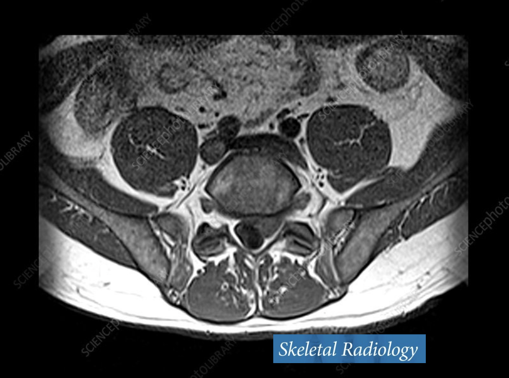
Dixon MRI can measure the proportion of fat within muscle. The difficulty is that each muscle first has to be carefully identified and separated from the surrounding tissue — a process known as segmentation. Until now, this has usually required time-consuming manual work.
We developed and tested an automated deep-learning method to measure fat in four key lumbar spine muscle groups: psoas, iliacus, quadratus lumborum, and the erector spinae and multifidus complex.
The method was trained using just 26 manually labelled MRI scans, then tested against expert manual measurements. It was then applied to MRI scans from 173 healthy adults, aged 20 to 70, ranging from sedentary volunteers to highly active recreational cyclists.
We found that the automated method closely matched manual measurement and produced new reference values for healthy levels of fat in lumbar spine muscles. The findings also showed that age and physical activity were important contributors to spinal muscle fat levels.
In short…
The study
Our objective was to develop and test an automated method for measuring fat in lumbar spine muscles from MRI scans.
We used Dixon MRI scans, which can measure fat fraction within muscle. 26 scans were manually labelled to train and test the deep-learning model.
The model measured fat in four lumbar spine muscle groups: psoas, iliacus, quadratus lumborum, and erector spinae plus multifidus.
The trained model was then applied to MRI scans from 173 healthy adults aged 20 to 70, including sedentary volunteers and recreational cyclists.
What we found
The automated method closely matched manual measurements of muscle fat.
It performed better than a previous multi-atlas segmentation method.
It produced healthy reference values for fat levels in lumbar spine muscles.
Fat levels varied between different muscles.
Age and physical activity were important factors in explaining differences in lumbar spine muscle fat.
What it means
This study shows that AI can make detailed muscle fat measurement much faster and more practical.
Faster automated measurement makes it more realistic to study large groups of people over time.
The new reference values can help future research into sarcopenia, spinal conditions and age-related muscle degeneration.
The method could also help clinicians and researchers monitor the effects of exercise, rehabilitation and other interventions on spinal muscle health.