How do core muscles differ between active and inactive people?

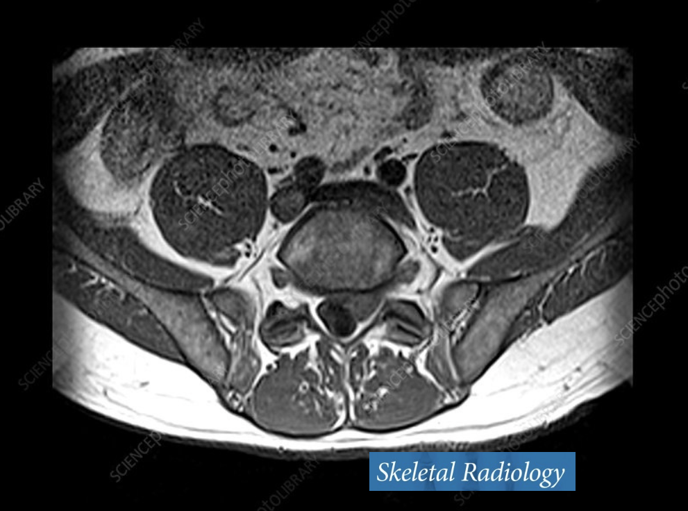
MRI cross-sectional image of lumbar spine. Automation enables us to compare large numbers of scans.
Published March 2026 in Skeletal Radiology
Core muscles play a central role in movement, stability and physical function. But measuring their health across multiple muscles is complex and time-consuming. In this study, we used an automated method to analyse MRI scans, comparing muscle fat and lean muscle volume in highly active and physically inactive adults, and developing new combined measures of overall core muscle health.
MRI can capture detailed information about muscle size and fat content. The challenge is processing those images: each muscle must be identified and measured individually, which has traditionally required slow, manual work.
Here, we applied an automated image analysis approach to Dixon MRI scans of the lumbar spine and pelvis. This enabled consistent measurement of fat fraction and lean normalised muscle volume across seven core muscles: psoas major, iliacus, quadratus lumborum, erector spinae/multifidus, and the three gluteal muscles.
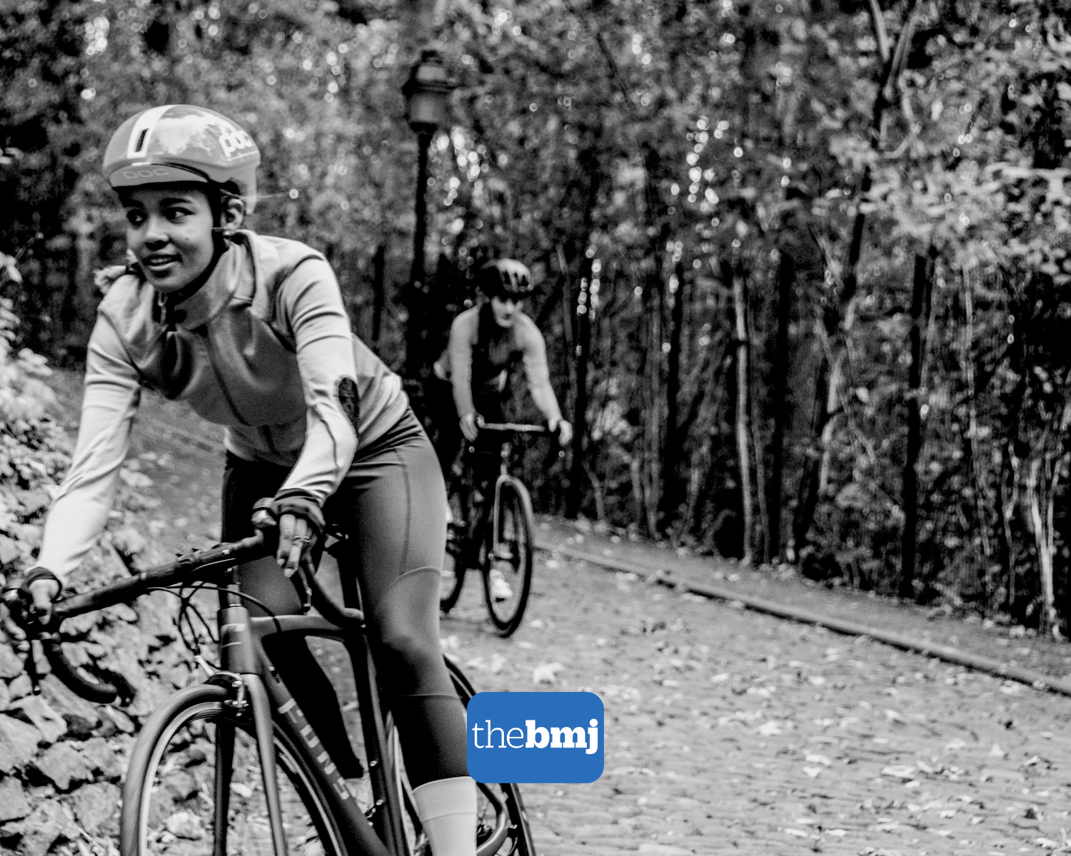
The study compared 84 highly active cyclists with 85 physically inactive adults. The automated approach also allowed us to generate combined “core scores” — summarising average fat fraction and total lean muscle volume across all muscles.
We found that cyclists had lower muscle fat and higher lean muscle volume than the inactive group across the muscles measured. Age was strongly associated with muscle fat, while activity level, sex and BMI were important predictors of lean muscle volume.
The study also shows that combined core scores can provide a useful overall measure of muscle health, capturing patterns that may be missed when looking at individual muscles alone.
In short…
The study
Our objective was to compare core muscle composition at different activity levels.
We analysed MRI scans using an automated image processing method.
We measured fat fraction and lean normalised muscle volume in seven core muscles.
We compared 84 highly active cyclists with 85 physically inactive adults.
We also created combined core scores for overall muscle fat and lean muscle volume.
What we found
Cyclists had lower muscle fat than physically inactive adults.
Cyclists had higher lean muscle volume across the muscles measured.
Age was strongly associated with increased muscle fat.
Activity level, sex and BMI were important predictors of lean muscle volume.
Combined core scores provided a clear summary of overall muscle composition.
What it means
Automated MRI analysis makes multi-muscle measurement faster and more consistent.
This makes large-scale and longitudinal studies more practical.
Physical activity is associated with healthier core muscle composition.
Looking across multiple muscles gives a more complete picture than focusing on one alone.
Combined core scores could support future research into ageing, sarcopenia and musculoskeletal health.